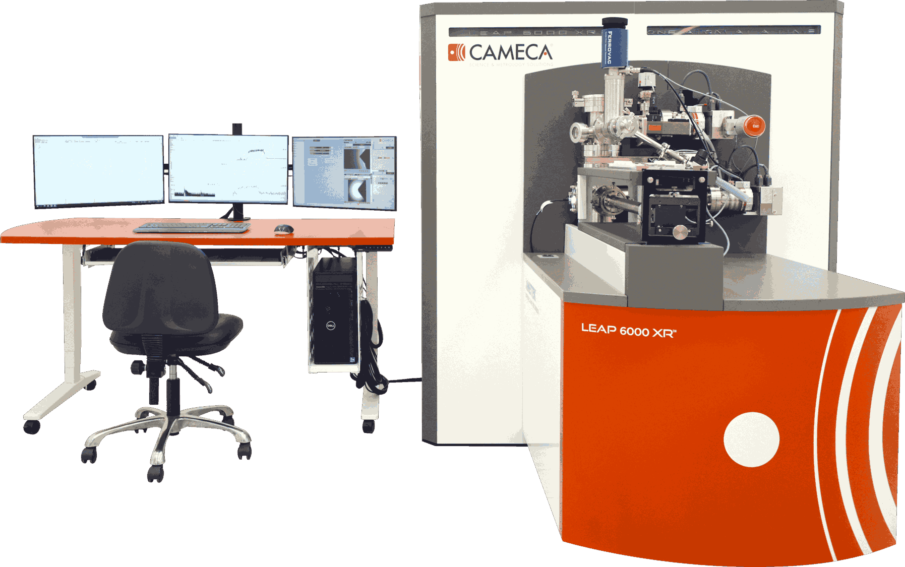
Atom Probe Tomography
Asset number
MC-SA-0007
Model
LEAP 6000XR
Brand
CEMACA
Asset Leader
邱嘉雯 Carmen Jiawen QIU
Contact email
Location
W1-122
Main specifications
1. Vacuum system of vacuum chamber: Level3, vacuum of the analysis chamber<1×10^-10 torr;
2. Max. voltage pulse frequency: 500 kHz;
3. Max. laser pulse frequency: 400kHz;
4. UV laser wavelength: 257.5nm; Finest focal spot:<3 µm;
5. Max. data collection rate ions/min: > 4M;
6. Time of Flight (TOF) mass spectrometer, ion flight path length: 382mm;
7. Mass resolution:
Mass resolution (half width): 1:1000
Mass resolution (one tenth of aspect ratio):1:475
Mass resolution (1% aspect ratio): 1:275;
8. The Vacuum-Cryogenic Transfer Module (VCTM) can provide a high vacuum and low temperature environment for sample transportation between the FIB-SEM and APT.
Main functions
Atom Probe Tomography is the only material analysis technique offering extensive capabilities for both 3D imaging and chemical composition measurements at the atomic scale (around 0.1-0.3nm resolution in depth and 0.3-0.5nm laterally). It is able to study the size, distribution, and composition of nano-scale microstructures (such as precipitates, clusters, and GP zones), as well as the segregation behavior at various internal interfaces (such as grain boundaries, phase boundaries, and interlayer interfaces in multilayer structures).
The working principle involves applying a high voltage to a needle-shaped sample, supplemented by electrical pulses or laser pulses, one or more atoms are evaporated from the surface, by field effect (near 100% ionization), and projected onto a Position Sensitive Detector with a very high detection efficiency.
This technique provides unique tomographic maps of elemental distribution at atomic level within small volumes of materials covering metals, semiconductor devices, nanomaterials, minerals, as well as organic and biological materials.
Precautions for sample testing
APT Sample Requirements:
1. Needle-shaped specimen.
2. Tip curvature radius of the specimen should be between 50 and 100 nm.
3. Region of interest should be located within 50–200 nm from the top of the specimen.
4. Specimen must be able to fit onto the APT sample holder (Puck).
Success rate and results of the test are closely related to sample preparation, please contact us to discuss the sample preparation requirements before proceeding.