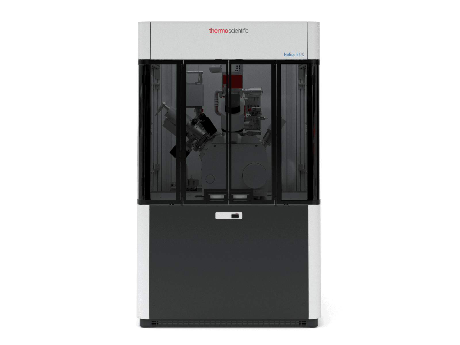
Dual-Beam Focused Ion Beam System
Asset number
MC-MP-0001
Model
Helios 5 UX
Brand
Thermo Fisher Scientific Brno s.r.o
Asset Leader
侯雨箫 Yuxiao HOU
Contact email
Location
W2-133
Main specifications
1. Bi ion source for analysis, ion beam current: >40 nA, maximum repetition rate: 50 kHz (for all modes of operation). Pulsed ion beam current: ≥ 40 pA, minimum pulsed beam diameter: ≤ 50 nm (for high-resolution imaging modes). Fast imaging mode available, minimum beam spot diameter: ≤ 90 nm @ 350 pA DC
2. G-SIMS analysis is supported in hardware and software, and G-SIMS analysis can be performed on organic macromolecules, and the G parameter can be changed in real time to change the molecular ion peak and fragment peak ratio, which helps to determine organic macromolecules.
3. Analytical sensitivity > 8.0×10⁸ Al+/nC @ 7000 (FWHM). Mass resolution when analyzing insulators (on PET: 104amu): ≥ 18000 (half-peak width; 100% transmission rate)
4. High-performance Orbitrap mass spectrometer with mass accuracy: < 1 ppm (RMS), enabling Ar ion cluster ion sources with minimum ion beam spot < 3um.
5. EDR Analyzer: maximum count rate > 1×10⁷ counts/sec in linear response range
Main functions
1. New Phoenix ion microscope cartridges offer excellent low voltage performance for the highest quality, spot-on, ultra-thin sample preparation for TEM and APT applications
2. AutoTEM 5 fully automated TEM sample preparation software for the fastest, easiest, fully automated, unattended TEM sample preparation and cross-sectional processing
3. Best Elstar Electron Microscope Cartridges with Smart Align and FLASH technology to obtain nano-scale information in the shortest time for users of any experience level
4. A new generation of High Current UC+ Monochromator Technology in the electron gun provides sub-nanometer resolution at low voltages, revealing the most detailed information, with an electron beam resolution of up to 0.7nm at 1KV
5. Up to 6 integrated in-barrel and under-lens detectors to capture high quality, sharp, charge-free images and provide the most complete sample information
6. Auto Slice&View 4 Software to precisely locate the region of interest, acquire the highest quality, multi-modal internal and 3D information, fully automated 3D slicing and imaging, and enable 3D EDS & EBSD reconstruction
7. Nano-Prototype Preparation for fast and accurate processing, etching and deposition of complex structures smaller than 10 nm
8. Highly accurate and stable 150 mm stroke piezoelectric ceramic driven sample stage and sample chamber Nav-Cam optical navigation camera for precise sample navigation